A Medical AI System That Reads Both X-Rays and CT Scans Natively
A single medical AI system now handles 2D images like X-rays and 3D scans like CTs — and board-certified radiologists ranked its reports highest against GPT-5.2 and Gemini-3-Flash.
- What they did — Built a multimodal medical AI system with a compositional vision encoder that natively processes both 2D images (X-rays, pathology slides) and 3D volumes (CT/MRI scans), plus a new evaluation framework that decomposes report quality into matched, missed, and hallucinated clinical findings.
- Key result — ClinFusion outperformed leading open-source medical MLLMs on 20 of 24 benchmarks and proprietary models like GPT-5.2 and Gemini-3-Flash on 13 of 16 multimodal benchmarks; six board-certified radiologists ranked its reports highest in a blinded evaluation of 300 clinical cases.
- Why it matters — For teams building medical AI systems, this suggests that composing multiple specialized vision encoders with guided fusion outperforms monolithic encoders — and that current report evaluation metrics poorly reflect what radiologists actually value.
The Problem
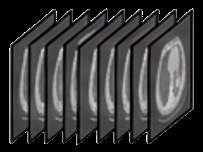
Medical imaging spans wildly different formats: flat 2D images like chest X-rays and pathology slides, and volumetric 3D scans like CTs and MRIs. Current medical AI models typically handle this diversity in one of two unsatisfying ways. The "2D-centric" approach treats everything as flat images by sampling slices from 3D volumes, which "inevitably sacrifices critical 3D structural information" [§1]. The "3D-centric" approach bolts on a separate 3D encoder but requires "extensive and complex alignment procedures" [§1].
Beyond the architecture problem, there's an evaluation problem. The standard way to judge AI-generated radiology reports — comparing generated text against a reference report using metrics like BLEU or BERTScore — is "fundamentally flawed" [§1]. Real radiologists don't write comprehensive reports in a vacuum; they interpret images guided by a patient's clinical context, focusing on specific anatomical regions. Current metrics also can't distinguish between a report that's accurate but incomplete versus one that's complete but hallucinates findings — a critical distinction in medicine [§1].
What They Did
ClinFusion tackles both problems with two main contributions: a new vision encoder architecture and a new evaluation framework.
**The vision architecture** replaces the typical single-encoder design with a compositional system. Think of it like assembling a diagnostic team rather than relying on one generalist. A foundational vision transformer (the generalist) provides a well-aligned base representation. Then an ensemble of specialist 2D encoders — each trained on different types of medical images — progressively enriches that base representation through what the authors call Cascade Spatial-Aware Locality (CaSL) Fusion [§1].
CaSL Fusion works in a cascaded, not parallel, fashion. Rather than averaging outputs from multiple encoders simultaneously (as prior work like Cambrian-1 does [§1]), each specialist encoder feeds its features into the next stage, progressively refining the representation while preserving the foundational encoder's alignment with the language model. For 3D data like CT scans, a dedicated 3D encoder is incorporated through "2D-anchored depth-aware CaSL Fusion," where the already-aligned 2D features serve as an anchor to guide the integration of 3D spatial information [§1].
**The evaluation framework** has two parts. First, MedIF-Bench tests whether medical AI models can follow complex clinical instructions — a capability that domain-specific fine-tuning often degrades, yet is "a prerequisite for any meaningful downstream evaluation and clinical deployment" [§1]. Second, a Region-of-Interest (RoI)-grounded evaluation method for report generation conditions on patient-specific clinical context (like which anatomical areas are clinically relevant) and uses an LLM judge to decompose each report into matched, missed, and hallucinated findings [§1]. This gives a factualness-driven breakdown rather than a single similarity score.
The system also includes an agentic tool-use layer that can invoke external specialist models for tasks like organ segmentation and retrieve current medical literature for citation-grounded answers [§1].
The Results
ClinFusion outperforms leading open-source medical MLLMs (including Hulu-Med and Lingshu) on 20 out of 24 benchmarks spanning visual question answering, report generation, instruction following, and text-only medical tasks [Abstract]. Against proprietary models, it demonstrates stronger multimodal capabilities than GPT-5.2 and Gemini-3-Flash on 13 out of 16 benchmarks [Abstract].
The most clinically meaningful validation comes from a blinded evaluation: six board-certified radiologists independently ranked reports from ClinFusion, Gemini-3-Flash, and Hulu-Med across 300 clinical cases covering CT and X-ray modalities. ClinFusion produced the highest-ranked reports across factual accuracy, completeness, and clinical utility [§1].
The evaluation framework itself was also validated: the RoI-grounded metric achieved "the strongest correlation with expert judgment among all eleven automatic evaluation metrics examined" [§1]. This is a notable finding on its own — it suggests that existing standard metrics for report generation are substantially misaligned with what radiologists actually care about.
Several limitations deserve attention. The benchmarks, while extensive at 24 tasks, represent controlled evaluation settings. Real clinical workflows involve mixed imaging protocols, incomplete patient histories, and edge cases that structured benchmarks don't capture. The radiologist evaluation, while blinded, covered 300 cases across only CT and X-ray modalities [§1] — pathology, fundus imaging, and MRI were not included in this expert validation. The agentic tool-use system, while showing "consistent improvements in both text-only and multimodal clinical scenarios" [§1], adds complexity and latency that the paper does not characterize for deployment contexts. And the system's performance against proprietary models was measured on 16 multimodal benchmarks, meaning it underperformed on 3 of them — the paper does not detail where it falls short.
Why It Matters
For builders working on medical AI systems, the cascaded encoder design offers a concrete architectural lesson: composing specialist encoders in sequence, anchored by a well-aligned foundation model, outperforms both monolithic encoders and parallel aggregation approaches. The code and models are publicly available on GitHub and HuggingFace [Abstract], making this directly testable.
For decision-makers evaluating medical AI capabilities — whether in hospital IT, regulatory bodies, or health-tech companies — the evaluation findings may matter more than the model itself. The demonstration that standard automated metrics poorly correlate with radiologist judgment [§1] should prompt skepticism toward any medical AI system evaluated solely on BLEU, ROUGE, or BERTScore. The RoI-grounded evaluation methodology, which decomposes reports into matched, missed, and hallucinated findings, provides a more trustworthy signal for procurement and validation decisions. If your organization is assessing AI-generated radiology reports, adopting factualness-driven decomposition metrics — rather than text-similarity scores — is actionable today.